Upward Pateller Fixation
Reprinted from: The Atlanta Equine Clinic
Intermittent upward patellar fixation is a condition whereby the horse’s pelvic limb temporarily "locks" in extension. As a result, there is a delay in flexion of the limb. The delay in flexion can range from milli-seconds to over several minutes. A short delay in flexion may manifest only as a subtle pelvic limb asymmetry or lameness; severely affected horses (with a long delay in flexion) may be unable to flex the affected limb without assistance.
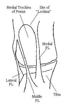
What is the "Patella"? The horse’s stifle joint is analogous to the human knee. Just like humans, horses have a patella, or "knee cap", which slides along the distal aspect of the femur (thigh bone) during flexion of the joint. The patella slides within a groove (called the trochlear groove) and serves as a fulcrum for the extensor muscles and their tendons as they course over the front of the stifle (or knee) joint. The patella is attached proximally to the quadriceps and biceps femoris muscles and distally to the tibia. In humans, the patella is attached to the tibia by one distal patellar ligament. Horses have 3 distal patellar ligaments: the medial patellar ligament, the middle patellar ligament, and the lateral patellar ligament.
How does the horse ‘lock’ the pelvic limb? Horses have the ability to lock (or fixate) the pelvic limb in extension. This is possible due to the unique anatomy associated with the horse’s stifle joint. The proximal aspect of the medial femoral trochlea is shaped similar to a hook or ski jump. By placing the space between the medial and middle patellar ligaments over this hook, horses can "lock" their pelvic limbs in extension. Once locked, minimal effort is required to maintain limb extension. A similar locking apparatus in the thoracic limbs allows horses to sleep while standing. Therefore, patellar fixation while standing is a normal process in the horse.
What is ‘intermittent upward patellar fixation’? Although patellar fixation is normal in the standing horse, it can produce pelvic limb dysfunction if it occurs during exercise. Inadvertent locking of the patella over the medial femoral trochlea prevents normal flexion of the affected limb(s). Consequently, pelvic limb asymmetry and lameness frequently become evident.
What causes upward patellar fixation? There are 3 primary causes of upward patellar fixation in the horse:
Lack of fitness: Lack of quadriceps and/or biceps femoris muscle tone results in an inability to quickly pull the patella up and off of the medial femoral trochlea.
Straight or upright pelvic limb conformation: This places the medial femoral trochlea further distad in closer proximity with the patella, facilitating patellar fixation.
Excessive distal patellar ligament length: This places the patella proximad in closer proximity with the medial femoral trochlea, where it can inadvertently "catch" or "lock"
It should be noted that the factors which cause upward patellar fixation are often interrelated. For example, an unfit horse will generally have increased laxity (and therefore increased length) of the distal patellar ligaments. Furthermore, if unfitness is secondary to another disease process (such as neurologic disease), intermittent upward fixation may also occur secondarily. Therefore, it is important to assess the horse as a whole prior to determining the cause for upward patellar fixation.
What are the clinical signs? Horses with intermittent upward patellar fixation will exhibit clinical signs during their attempt to flex the pelvic limb from an extended position. In acute severe cases, the pelvic limb may stay locked in extension. The horse may not be able to flex the stifle and tarsus without assistance. In some instances, the condition may temporarily resolve only to recur after taking a few steps. These signs are quite obvious and diagnosis is relatively simple if the condition is severe. Most of the time, however, there is only a "catching" of the patella as it slides up and over the hook and the limb does not truly lock in extension. In this situation, there may only be a mild pelvic limb asymmetry or lameness. This type of lameness can be easily confused with other problems and therefore may present a dilemma in regard to accurate diagnosis. Following are common clinical signs associated with mild to moderate forms of intermittent upward patellar fixation:
Non-weightbearing pelvic limb lameness
This may be distinguished from tarsal (hock) soreness which is usually weightbearing in nature
The horse will frequently drag the toe of the affected limb(s) during exercise
Visible wearing of the dorsal aspect of the toe/shoe may be apparent.
The foot of the affected limb(s) will have a low-arc flight pattern
The horse will usually exhibit a shortened cranial phase to the stride
Resistance in the canter
The horse will resist the canter, particularly if circled toward the more affected limb
Resistance may be most noticeable during the transition between the trot and canter, when the horse is forced to extend the pelvic limb for a prolonged period
Many horses will toss their head, rear, or stop when asked to canter. This may be due to their "anticipation" of impending upward patellar fixation.
The horse would rather trot than canter (which is harder for the normal horse)
Consistent lead changes or cantering on the wrong lead
The horse avoids prolonged pelvic limb extension with the affected limb. This is particularly apparent when cantering in a circle towards the affected limb.
The canter is very rough or "bouncy"
This occurs as a result of consistent delay in pelvic limb flexion from the extended position
Swelling, heat, and/or pain may be associated with one or both stifle joints
Upward patellar fixation causes patellar instability which in turn may result in femoropatellar synovitis
The horse drags his hind toes during exercise
Resistance and/or difficulty when walking up and down hills, or when backing up
These situations force the horse to extend the pelvic limb for a prolonged period
Rather then fully extend the pelvic limb(s), the horse may "crouch" while walking
Rather than flex the pelvic limb(s) normally, horses will often swing their limbs to the outside
This may cause the lameness to be confused with neurologic disease (such as EPM or stringhalt)
Lameness is most severe when the horse is first taken out of the stall
Many horses will improve as the workout progresses
Lameness becomes more obvious following an extended period of stall rest
Loss of muscle and patellar ligament tone exacerbate the upward patellar fixation
The horse does not improve (and may worsen) as a result of taking time off
The horse does not respond to anti-inflammatory (e.g. Phenylbutazone) therapy
Intermittent upward patellar fixation is a mechanical problem and is not inflammatory-mediated
As with many cases of pelvic limb lameness, secondary abnormalities such as thoracolumbar ebaxial (back) and proximal thoracic suspensory ligament soreness are also present. These are generally detected during the passive lameness evaluation and are suggestive of chronic pelvic limb asymmetry/ lameness.
How is upward patellar fixation diagnosed? Clinical signs are characteristic and, if the limb is locked in extension (i.e. the case is severe), diagnosis is simple. As previously mentioned, however, most cases are mild and diagnosis may be more difficult. A detailed history and careful clinical evaluation are essential parts of a proper workup. One helpful diagnostic aid involves placing the horse in one or more situations where prolonged pelvic limb extension is normally required. Such situations include walking up and down hills, the trot-to-canter transition, and backing up. When confronted with these situations, the affected horse will either 1) demonstrate upward patellar fixation by temporarily locking the pelvic limb, or 2) cheat by switching leads, swinging the limbs to the outside, avoiding pelvic limb extension, etc.
Many times, a slight hitch or "catch" is visible as the pelvic limb begins to flex from an extended position. This "catch" is most easily detected by visualizing the point of the hock as the horse picks the limb up to advance it cranially. Infrequently, an audible "snap" or popping sound is also evident during exercise (particularly walking).
In many instances, upward patellar fixation can be produced in affected horses by manually forcing the patella upward and outward. The examiner may actually be able to keep the pelvic limb locked in extension using minimal effort.
Since the problem is usually secondary to conformation and/or level of fitness, it is almost always bilateral. However, affected horses historically exhibit clinical signs in one pelvic limb. It is not until the more affected limb is successfully treated that a problem in the contralateral limb is manifested.
How is upward patellar fixation treated? Currently, there are 5 forms of treatment for intermittent upward patellar fixation:
Exercise: Lack of fitness results in decreased thigh muscle and patellar ligament tone. With decreased supporting muscle and ligament tone, it becomes easier for the patella to lock on the femur and harder for it to replace within the trochlear groove. In subtle cases of upward patellar fixation where conformation is relatively good, increased exercise alone may result in resolution of the problem.
We frequently ask the client to grade the level of their horse’s current level of fitness on a scale of 1 to 10 (1=very unfit; 10=extremely fit). We suggest achieving a fitness level of at least 7-8 (if possible) prior to pursuing other forms of treatment. This will rule out unfitness as a major contributor to the problem as well as increase the effect of other therapy.
Corrective Shoeing: Since fixation of the patella occurs when the pelvic limb is extended, prolonging the extension phase of the stride can make "unlocking" more difficult. Alternatively, shortening the amount of time the pelvic limb spends in extension allows the horse to unlock his/her patella before the distal patellar ligaments become excessively tight. Since the conformation of the distal pelvic limb and/or the toe length is intimately related to pelvic limb breakover, the farrier can frequently alleviate the problem via corrective trimming/shoeing. Rolling and/or rockering the toe of the shoe, applying a full (egg-) bar shoe, and/or the use of wedged pads (when needed) are commonly used techniques. In many cases, we are able to help the pelvic limbs break over before intermittent upward patellar fixation occurs.
Hormonal Therapy: The administration of estrogen has shown to prove benefical for some horses exhibiting intermittent upward patellar fixation. The presence of estrogen within the body of the horse may increase tension of various supporting ligaments. These include the collateral, suspensory, cruciate, and distal patellar ligaments. Increasing distal patellar ligament tension helps to relocate the patellar further distad, thereby making upward patellar fixation more difficult. This in turn may alleviate clinical signs.
It should be noted that estrogen is also a powerful behavior modificator in the horse. It is often used for stallions and geldings that are excessively difficult to handle, aggressive towards people or other horses, or overly anxious at shows and other events. Estrogen is very effective at reducing anxiety and resistance as well as improving overall behavior in these horses. Treatment usually consists of 2 injections of estrogen (25mg) in the muscle twice weekly for 4 consecutive weeks, then as needed therafter.
Administration of estrogen to mares usually causes them to exhibit clinical signs of estrus (heat). Since this change in behavior is generally undesirable, we do not recommend its use in mares.
Intraligamentous Infusion of Counterirritant: This form of therapy is usually referred to as "blistering". Blistering involves the inject of an irritative substance into soft tissue(s) in an attempt to create an inflammatory reaction. The irritative substance usually consists of iodine 2% in an almond oil base. This substance can elicit an inflammatory response for up to 30 days depending on the amount used and the location of injection. It is important to remember that fibrosis and scar tissue formation within normal soft tissues will occur as a result of severe inflammation. As you know, scar tissue does not function like normal soft tissue. Therefore, blistering in certain areas may inhibit proper function of associated soft tissue. It is for this reason that The Atlanta Equine Clinic typically does not institute blistering as typical form of treatment for soft tissue problems.
However, in the case of intermittent upward patellar fixation, we gain a biomechanical advantage by replacing normal tissue with scar tissue. The infusion of counterirritant within and around the medial and middle patellar ligaments results in the elicitation of an intense inflammatory reaction by the horse’s body. With inflammation, fibrosis and scarring of the patellar ligaments occur. During the scarring process, soft tissues will contract (shorten). As the patellar ligaments shorten, the patella is pulled up and over the hook of the medial femoral trochlea and into its normal position within the trochlear groove. At this point, it becomes more difficult for the horse to lock the patella and easier to flex the pelvic limb from an extended position. In our hands, this from of treatment has been extremely effective in a vast majority of cases involving intermittent upward patellar fixation.
Medial Patellar Desmotomy: The medial patella ligament is one of the key structures (along with the patella and middle patellar ligament) that is required to lock the patella on the femur. Since the problem represents the horse’s inability to quickly disengage the patella from the medial femoral trochlea, surgical resection of the medial patellar ligament results in complete resolution of the problem. Once the medial patellar ligament is resected, upward patellar fixation becomes impossible and the clinical signs associated with this condition disappear. Consequently, this has become a very popular form of treatment for horses with intermittent upward patellar fixation.
It is extremely important to note, however, that the medial patellar ligament also performs another function: stabilization of the patella within the trochlear groove of the femur. Without tension from the medial patellar ligament, the patella becomes unstable within the femoropatellar joint. Femoropatellar synovitis and frequently osteoarthritis result. Since the stifle is high-motion in nature, chronic inflammation within this joint poses a significant concern in regard to future performance soundness. Persistent femoropatellar joint inflammation typically needs to be addressed on a continual basis and often requires considerable maintenance therapy. It is for this reason that The Atlanta Equine Clinic views this form of treatment inappropriate except for the most severe of cases that have proven refractory to the other forms of therapy.